Category Archives: Uncategorized

Weed and Stress

In arecent blog entry, I wrote about how some of the young people I treat in my practice are using cannabis as a means of avoiding the anxiety-provoking stressors of becoming an adult and how that avoidance is contributing to a delay in maturation.

The good news is that most young people who experiment with cannabis in adolescence will not develop psychosis. But a small percent, especially with genetic loading, can develop psychotic symptoms at a younger age with exposure to cannabis.

In this entry, I want to explore a far more serious question: Does cannabis increase the chances of developing psychosis?

The answer is not a simple one. Cannabis appears to interact with neurodevelopmental processes in a way that in some people can contribute to the genesis of psychotic illnesses such as schizophrenia. However, fortunately for most people, this will not be the case. Global epidemiology studies attribute only a tiny amount of the risk of developing schizophrenia to the use of cannabis. (6) But even if this percentage is small, even as low as 1% of users, the increasing prevalence of cannabis use among young people, coupled with the appearance of relative safety (an image of safety that has been promoted by the cannabis industry, just as the safety of tobacco was once purported by the likes of R.J. Reynolds) and growing societal acceptance of cannabis means that more young people will be exposed to cannabis and this potential attendant harm. Even if this risk is relatively small, the level of disability and suffering associated with schizophrenia behooves us to work towards preventing any cases that we can.

Schizophrenia is a complex illness, likely resultant from disrupted neurotransmission, both on the cellular and network level. It typically emerges in late adolescence and early adulthood, before the age of 25. This is also the time when many people experiment with substance use, and cannabis is the most widely used substance among young people after alcohol and tobacco. In one study, 7.3% of people 12 and older reported using cannabis in the previous month compared with 52.1% who reported alcohol use and 26.7% who reported tobacco use. (1)

We clinicians have all seen the tragedy of young patients who, just as they are getting ready to launch into the world, began to develop the disabling symptoms of schizophrenia. Some of them have also been using cannabis. The question often arises from family members and patients: Did the cannabis cause the illness?

The simple answer in clinical practice is “we will never know,” because of course, there is no ability to have a “control” patient who is not exposed to cannabis to see if the illness would not develop in its absence. Additionally, patients with schizophrenia and their families can better direct their efforts towards helping their loved one heal and manage their illness, rather than feeling regrets about past choices.

However, as clinicians, it is important for us to educate our young patients about the risks of psychosis associated with early, heavy cannabis use. Epidemiologic data indicates that young people who are daily users of cannabis before the age of 15 have a greater risk of developing schizophrenia than their peers who either abstain from cannabis or wait until adulthood to begin using the drug. This risk is further increased by the use of cannabis that is high in THC (9-delta tetrahydrocannabinol), the psychoactive ingredient in cannabis, as many contemporary strains are grown for high levels of this compound. (2) This relationship between psychosis and cannabis use appears to be dose dependent, which further underscores the harm reduction message that while no use is bestany reduction or forestalling of use is better. (3)

Curiously, subjects with prodromal schizophrenia have been found to have elevated endocannabinoid activity. Anandamide, the endogenous ligand for the cannabinoid-1 receptor, has been found to be elevated up to eight times normal in the CSF of patients with emerging schizophrenia. This may represent an attempt by the brain to restore homeostasis to a brain that has become overactive with dopamine. Anandamide may be trying to “put the brakes on” the emerging excessive dopamine activity seen in emergent schizophrenia. Unfortunately, exposing a brain that is already struggling with dopamine homeostasis to the perturbing activity of THC may further worsen the psychotic symptoms. (7)

Why might cannabis use both increase the risk of psychosis? It may be partially explained by genetics. The gene that codes for COMT (catechol-O-methyltransferase) has some regulation of dopamine degradation at the synapse. Schizophrenia is associated with an excess of dopamine activity, particularly at the mesolimbic tract of the brain. (4) Carriers of the Val158Val polymorphism for the COMT gene were more likely to develop psychotic symptoms  at a younger age after exposure to cannabis than peers who carried the Met158Met polymorphism. (5) As genetic testing becomes cheaper, might it make sense to screen young people prior to the age at which drug use is typically initiated for this polymorphism and warn those who are Val/Val carriers that they are at a higher risk for developing a psychotic illness with cannabis exposure?

So what do we tell our young patients? One – by first acknowledging that we have damaged our credibility with a historic message that “all drugs are bad for all people all the time,” young users may be more willing to hear a more nuanced message. Two – there are real risks for young people who use cannabis daily. If they haven’t tried the drug, try to explain the benefits of waiting. Each year that passes before drug initiation lessens the risk of developing psychosis. If they have used the drug, and are unwilling to stop, adopt a position of harm reduction: any reduction of use is better than more use. Discuss changing the relationship with the drug to use less of it and to use it less frequently.

References

1. Results from the 2012 National Survey on Drug Use and Health: Summary of National Findings. Substance Abuse and Mental Health Services Administration.

2. Di Forti M, Sallis H, Allegri, F, et al. Daily use, especially of high-potency cannabis, drives the earlier onset of psychosis in cannabis users. Schizophr Bull. 2014;40(6):1509-17.

3. Andreasson S, et al. Lancet. 1987;2(8574):1483-1486. Fergusson DM, et al. Addiction. 2005;100(3):354-366. Henquet C, et al. BMJ. 2005;330(7481):11. Moore TH, et al. Lancet. 2007;370(9584):319-328. Schubart CD, et al. Acta Psychiatr Scand. 2011;123(5):368-375.

4. Stahl, S  (2013) Stahl’s Essential Psychopharmacology. Cambridge Press

5. Estrada G, et al. Acta Psychiatr Scand. 2011;123(6):485-492. Verdejo-Garcia A, et al. Neuropsychopharmacology. 2013;38(8):1598-1606.

6. Degenhardt L, Ferrari A, Calabria B, et al. The global epidemiology and contribution of cannabis use and dependence to the global burden of disease: results from the GBD 2010 study. PLoS One. 2013;8(10):e76635. doi:10.1371/journal.pone.0076635. 

7. Leweke FM. Curr Pharm Des. 2012;18(32):5188-5193. Meltzer HY, et al. Am J Psychiatry. 2004;161(6):975-984. Leweke FM, et al. Transl Psychiatry. 2012;2:e94. Guffrida A, et al. Neuropsychopharmacology. 2004;29(11):2108-2114.

Andrew Penn was trained as an adult nurse practitioner and psychiatric clinical nurse specialist at the University of California, San Francisco. He is board certified as an adult nurse practitioner and psychiatric nurse practitioner by the American Nurses Credentialing Center. Currently, he serves as an Assistant Clinical Professor at the University of California-San Francisco School of Nursing. Mr. Penn is a psychiatric nurse practitioner with Kaiser Permanente in Redwood City, CA, where he provides psychopharmacological treatment for adult patients and specializes in the treatment of affective disorders and PTSD. He is a former board member of the American Psychiatric Nurses Association, California Chapter, and has presented nationally on improving medication adherence, emerging drugs of abuse, treatment-resistant depression, diagnosis and treatment of bipolar disorder, and the art and science of psychopharmacologic practice.

Exercise and Mental Health

New Guidelines: Exercise Key Part of Mental Health Treatment

October 15, 2018


New guidelines from the European Psychiatric Association promote exercise as a core part of treatment for severe mental illnesses, including major depressive disorder and schizophrenia. The recommendations follow a review of existing research on the effect of physical activity on mental health disorders.

Findings from the meta-review and the resulting position statement and guidelines were published online in European Psychiatry.

“Our comprehensive review provides clear evidence that physical activity has a central role in reducing the burden of mental health symptoms in people with depression and schizophrenia,” said lead investigator Brendon Stubbs, PhD, King’s College London, and South London and Maudsley NHS Foundation Trust, London, United Kingdom.

Prescribing Exercise as an Intervention for Depression

“Our guidelines provide direction for future clinical practice. Specifically, we provide convincing evidence that it is now time for professionally-delivered physical activity interventions to move from the fringes of healthcare and become a core component in the treatment of mental health conditions.”

According to the analysis, moderate-intensity aerobic exercise 2 to 3 times a week, for a total of 150 minutes, eases symptoms of depression and schizophrenia. In people with schizophrenia spectrum disorders, such physical activity also improves cognition and cardiorespiratory health. Combining aerobic with resistance exercise, according to evidence, may also improve outcomes in people with major depression and schizophrenia spectrum disorders.

To better support the treatment of patients with severe mental illness, new association guidelines advocate for structural changes to mental health facilities to include exercise facilities and exercise specialists, as well as updated insurance reimbursement guidelines that support the recommended physical activity interventions.

Trivia: How Much Exercise Is Needed to Prevent Depression?

“Our systematic review of top-tier evidence has convincingly demonstrated that exercise training, delivered by physical activity professionals, does indeed provide an effective add-on treatment for improving both physical and mental health outcomes in people with mental illness,” Dr. Stubbs said.

The International Organization of Physical Therapists in Mental Health also endorsed the guidelines.

—Jolynn Tumolo

References

Stubbs B, Vancampfort D, Hallgren M, et al. EPA guidance on physical activity as a treatment for severe mental illness: a meta-review of the evidence and Position Statement from the European Psychiatric Association (EPA), supported by the International Organization of Physical Therapists in Mental Health (IOPTMH). European Psychiatry. 2018 October 1;[Epub ahead of print].

European Psychiatric Association (EPA) comprehensive review demonstrates that exercise is an effective treatment for major mental health conditions and should form a core part of treatment [press release]. Philadelphia, Pennsylvania: Elsevier; October 1, 2018.

Donald Rauh M.D., Ph.D., FAPA
Diplomate of the American Board of Psychiatry & Neurology
Board Certified in General Psychiatry and in  Child & Adolescent Psychiatry

Cured Meats and Mania

Eating Cured Meats May Increase Risk for Mania

Peter Roy-Byrne, MD reviewing Khambadkone SG et al. Mol Psychiatry 2018 Jul 18.
A specific association between ingestion of nitrate-cured meat products and mania is bolstered by behavioral and genetic findings in animals.
Although there is a significant genetic contribution to bipolar illness, environmental factors, including diet, are thought to be important. These researchers surveyed the dietary history (foods “ever eaten”) of a broad group of psychiatric patients receiving hospital or outpatient care (bipolar mania, 217 individuals; bipolar depression, 91; major depression, 79; schizophrenia, 371) and 343 healthy controls.
A history of eating nitrate-cured meat products specifically was associated with being in the mania group. A subgroup of 42 patients with mania and 35 controls were asked about the type of cured meat products eaten; meat sticks, beef jerky, and turkey jerky were specifically implicated.
In follow-up experimental studies of rats, a meat diet with added nitrates produced both overall hyperactivity and hyperactivity to novel stimuli. The diet was also associated with expressed gene dysregulation involving serotonin, nuclear factor signaling, and sphingosine-1-phosphate signaling and with increases in small-bowel bacteria species previously linked to cognitive and behavioral alterations in animals.
Donald Rauh M.D., Ph.D., FAPA
Diplomate of the American Board of Psychiatry & Neurology
Board Certified in General Psychiatry and in  Child & Adolescent Psychiatry

Heavy Metals found in some Baby Foods

Consumer Reports, an independent, nonprofit organization that works side by side with consumers to create a fairer, safer, and healthier world, writes regarding our recently completed testing of baby and toddler food for elements commonly known as “heavy metals,” including cadmium, lead, and inorganic arsenic. Based on our findings, we urge the Food and Drug Administration (FDA) to take several steps to protect the public from these potentially harmful contaminants in the food supply.

As the FDA is aware, exposure to even small amounts of these elements may, over time, increase the risk of several serious health problems, including those involving carcinogenic, cognitive, and reproductive effects. Babies and young children are the most at risk, particularly given the potential harms of heavy metal exposure on developing brains in the form of lower IQ and behavior problems.

As we report in a major story published today on CR.org, Consumer Reports’ food safety team analyzed 50 nationally distributed foods made for babies and toddlers, including baby cereals, packaged fruits and vegetables, packaged entrées, and packaged snacks. We found:

  • Every product had measurable levels of at least one of these heavy metals: cadmium, lead, or inorganic arsenic.
  • Around two-thirds (68%) had worrisome levels of at least one heavy metal.
  • Among the 50 foods tested, 15 would pose potential health risks to a child regularly eating just one serving or less per day.
  • Snacks and products containing rice and/or sweet potatoes were particularly likely to have high levels of heavy metals.
  • Certified organic foods were as likely to contain heavy metals as conventional ones.

We are concerned by these results, especially because a recent nationwide survey we conducted of over 3,000 respondents found misconceptions among parents about the safety of baby food. Four in 10 of the parents we surveyed believe children’s packaged foods are lower in heavy metals than other packaged foods. This is despite our testing that showed worrisome levels in many children’s food products. About half of respondents think baby food is already regulated more stringently than other packaged food, and an even larger 72% think it should be.

Accordingly, as a part of our published story, Consumer Reports provides several recommendations to parents. We lay out several steps parents can take to reduce heavy metals exposure, including to eat a broad array of healthful whole foods; limit intake of infant rice cereal and packaged snacks and be conscious of intake of fruit juices; and to choose rice that generally contains less inorganic arsenic. We also encourage them not to panic, and to remember that significant exposure to heavy metals does not guarantee that a child will have health problems, merely that it increases the chance of them.

As parents seek to reduce children’s exposure to heavy metals and its associated risks, they should not be left to fend for themselves. Food companies and the FDA have essential roles to play, particularly given another, more encouraging finding of ours: among the 50 products tested, 16 had less concerning levels of the heavy metals. Indeed, for some products, there were some of the metals that were not measurable at all. This suggests that baby food manufacturers across the industry should be able to achieve similar results. We encourage these companies to implement strong, recognized best practices—such as the Codex Alimentarius Code of Practice for the Prevention and Reduction of Lead Contamination in Foods—and urge the FDA to ensure they do so. We appreciate the important, ongoing work of the FDA’s Toxic Elements Working Group in this area, and recognize the agency’s ability to take enforcement action on a case-by-case basis and its efforts to prevent contaminated food from being imported.

However, we also recognize that the FDA has no specific limits in place to restrict contaminants in the vast majority of children’s food. With the agency’s own data and the work of Consumer Reports and other public interest groups in mind, it is critically important for the FDA to take the following additional steps to protect the public and assist parents nationwide:

  • Establish aggressive targets. Because there are no established safe levels of heavy metals like lead, we urge the FDA to set a goal of having no measurable amounts of cadmium, lead, or inorganic arsenic in baby and children’s food—and to use the most sensitive testing methods to determine the presence of those elements.
  • Create and enforce benchmarks. To limit heavy metals in baby and children’s food, the FDA should set incremental targets for industry to meet along the way, while continually recognizing that the end goal must be always to have no measurable amounts. The agency also should insist that manufacturers meet strong, recognized best practices as described above.
  • Finalize proposed guidelines. By the end of 2018, the agency’s planned deadline, the FDA should finalize its guidances limiting inorganic arsenic in apple juice to 10 ppb, and limiting inorganic arsenic in infant rice cereal to 100 ppb. Also, it should revise existing guidance for lead in fruit juice to reduce the limit from 50 to 5 ppb, the standard for bottled water.

We appreciate your consideration of these critical issues, and kindly request your response to this letter at your earliest convenience. Consumer Reports looks forward to working with you to prevent contamination, reduce levels of heavy metals in food made for babies and children, and limit the risk of food safety harm to consumers nationwide.

For the full letter, click here.

Author

Jean Halloran and James Rogers

DNA testing

STAT (9/28, Robbins) reported that “several dozen companies” are now “probing patients’ DNA in search of insights to help inform decisions about what psychiatry medications patients should take,” and are even “touting applications for depression, bipolar disorder, attention deficit hyperactivity disorder, and post-traumatic stress disorder.
” Now, “some top psychiatrists say the evidence doesn’t support the commercial rush.” In fact, in a review published online April 25 in the American Journal of Psychiatry, “a task force of the American Psychiatric Association’s research council concluded that such genetic testing is not ready for prime time in their field.” The members of the task force wrote, “Although some of the preliminary published data sound promising…there is insufficient evidence to support widespread use of combinatorial pharmacogenetic decision support tools at this point in time.”
Donald Rauh M.D., Ph.D., FAPA
Diplomate of the American Board of Psychiatry & Neurology
Board Certified in General Psychiatry and in  Child & Adolescent Psychiatry

Brain Development and Preschoolers

Atypical brain development observed in preschoolers with ADHD symptoms

NIH-funded study uses high-resolution brain scans to uncover structural changes.

Children as young as 4 years old with symptoms of attention deficit hyperactivity disorder (ADHD) may have significant differences in brain structure, compared to children without such symptoms, according to researchers funded by the National Institutes of Health.

Their study is the first comprehensive examination of brain structure changes in preschoolers with signs of ADHD, a disorder marked by a pattern of inattention, hyperactivity and impulsive behavior. The study appears in the Journal of the International Neuropsychological Society.

“Researchers were expecting to find the beginning signs of possible atypical brain development,” said James A. Griffin, Ph.D., deputy chief of the Child Development and Behavior Branch at NIH’s Eunice Kennedy Shriver National Institute of Child Health and Human Development, which supported the research. “What they found in their high-resolution scans was solid evidence that these structural changes already are prominent by age 4 or 5.”

Previous studies have documented brain differences in adolescents with ADHD. However, few studies have looked for such differences in preschoolers, despite research citing ADHD as the most commonly diagnosed psychological disorder among young children.

The research, led by E. Mark Mahone, Ph.D., was conducted at the Kennedy Krieger Institute in Baltimore. The study included 90 young children: 38 typically developing preschoolers and 52 preschoolers with symptoms of ADHD.

The children’s scans revealed that those with ADHD symptoms had multiple areas with less brain matter volume than their typical peers, and these differences were consistent with parent reports of hyperactive and impulsive behaviors.

The researchers cited challenges collecting data, mainly getting youngsters to lie still during the brain scan, particularly children with ADHD-associated behavior. It is possible that the children who were eventually scanned had more moderate symptoms and, therefore, were better suited to participate in the study. The authors speculated that children with more severe ADHD may have more pronounced brain differences.

The research offers a greater understanding of brain development in children with ADHD symptoms, Griffin said. Researchers will continue to follow the children, monitoring brain changes or differences as the they grow older. The study provides the groundwork for future analysis of structural and functional brain changes in ADHD, which the researchers hope will provide new insights into how symptoms of the disorder relate to differences in the brain.

About the Eunice Kennedy Shriver National Institute of Child Health and Human Development (NICHD): NICHD conducts and supports research in the United States and throughout the world on fetal, infant and child development; maternal, child and family health; reproductive biology and population issues; and medical rehabilitation. For more information, visit https://www.nichd.nih.gov.

About the National Institutes of Health (NIH): NIH, the nation’s medical research agency, includes 27 Institutes and Centers and is a component of the U.S. Department of Health and Human Services. NIH is the primary federal agency conducting and supporting basic, clinical, and translational medical research, and is investigating the causes, treatments, and cures for both common and rare diseases. For more information about NIH and its programs, visit www.nih.gov.

NIH…Turning Discovery Into Health®

Reference

Jacobson, L.A., et al. Anomalous Brain Development is Evident in Preschoolers with Attention Deficit Hyperactive Disorder. Journal of the International Neuropsychology Society. 2018; doi:10.1017/S1355617718000103

Schizophrenia

Overview

Schizophrenia is a serious mental disorder in which people interpret reality abnormally. Schizophrenia may result in some combination of hallucinations, delusions, and extremely disordered thinking and behavior that impairs daily functioning, and can be disabling.

People with schizophrenia require lifelong treatment. Early treatment may help get symptoms under control before serious complications develop and may help improve the long-term outlook.

Symptoms

Schizophrenia involves a range of problems with thinking (cognition), behavior or emotions. Signs and symptoms may vary, but usually involve delusions, hallucinations or disorganized speech, and reflect an impaired ability to function. Symptoms may include:

  • Delusions. These are false beliefs that are not based in reality. For example, you think that you’re being harmed or harassed; certain gestures or comments are directed at you; you have exceptional ability or fame; another person is in love with you; or a major catastrophe is about to occur. Delusions occur in most people with schizophrenia.
  • Hallucinations. These usually involve seeing or hearing things that don’t exist. Yet for the person with schizophrenia, they have the full force and impact of a normal experience. Hallucinations can be in any of the senses, but hearing voices is the most common hallucination.
  • Disorganized thinking (speech). Disorganized thinking is inferred from disorganized speech. Effective communication can be impaired, and answers to questions may be partially or completely unrelated. Rarely, speech may include putting together meaningless words that can’t be understood, sometimes known as word salad.
  • Extremely disorganized or abnormal motor behavior. This may show in a number of ways, from childlike silliness to unpredictable agitation. Behavior isn’t focused on a goal, so it’s hard to do tasks. Behavior can include resistance to instructions, inappropriate or bizarre posture, a complete lack of response, or useless and excessive movement.
  • Negative symptoms. This refers to reduced or lack of ability to function normally. For example, the person may neglect personal hygiene or appear to lack emotion (doesn’t make eye contact, doesn’t change facial expressions or speaks in a monotone). Also, the person may have lose interest in everyday activities, socially withdraw or lack the ability to experience pleasure.

Symptoms can vary in type and severity over time, with periods of worsening and remission of symptoms. Some symptoms may always be present.

In men, schizophrenia symptoms typically start in the early to mid-20s. In women, symptoms typically begin in the late 20s. It’s uncommon for children to be diagnosed with schizophrenia and rare for those older than age 45.

Symptoms in teenagers

Schizophrenia symptoms in teenagers are similar to those in adults, but the condition may be more difficult to recognize. This may be in part because some of the early symptoms of schizophrenia in teenagers are common for typical development during teen years, such as:

  • Withdrawal from friends and family
  • A drop in performance at school
  • Trouble sleeping
  • Irritability or depressed mood
  • Lack of motivation

Compared with schizophrenia symptoms in adults, teens may be:

  • Less likely to have delusions
  • More likely to have visual hallucinations

When to see a doctor

People with schizophrenia often lack awareness that their difficulties stem from a mental disorder that requires medical attention. So it often falls to family or friends to get them help.

Helping someone who may have schizophrenia

If you think someone you know may have symptoms of schizophrenia, talk to him or her about your concerns. Although you can’t force someone to seek professional help, you can offer encouragement and support and help your loved one find a qualified doctor or mental health professional.

If your loved one poses a danger to self or others or can’t provide his or her own food, clothing or shelter, you may need to call 911 or other emergency responders for help so that your loved one can be evaluated by a mental health professional.

In some cases, emergency hospitalization may be needed. Laws on involuntary commitment for mental health treatment vary by state. You can contact community mental health agencies or police departments in your area for details.

Suicidal thoughts and behavior

Suicidal thoughts and behavior are common among people with schizophrenia. If you have a loved one who is in danger of attempting suicide or has made a suicide attempt, make sure someone stays with that person. Call 911 or your local emergency number immediately. Or, if you think you can do so safely, take the person to the nearest hospital emergency room.

Causes

It’s not known what causes schizophrenia, but researchers believe that a combination of genetics, brain chemistry and environment contributes to development of the disorder.

Problems with certain naturally occurring brain chemicals, including neurotransmitters called dopamine and glutamate, may contribute to schizophrenia. Neuroimaging studies show differences in the brain structure and central nervous system of people with schizophrenia. While researchers aren’t certain about the significance of these changes, they indicate that schizophrenia is a brain disease.

Risk factors

Although the precise cause of schizophrenia isn’t known, certain factors seem to increase the risk of developing or triggering schizophrenia, including:

  • Having a family history of schizophrenia
  • Increased immune system activation, such as from inflammation or autoimmune diseases
  • Older age of the father
  • Some pregnancy and birth complications, such as malnutrition or exposure to toxins or viruses that may impact brain development
  • Taking mind-altering (psychoactive or psychotropic) drugs during teen years and young adulthood

Complications

Left untreated, schizophrenia can result in severe problems that affect every area of life. Complications that schizophrenia may cause or be associated with include:

  • Suicide, suicide attempts and thoughts of suicide
  • Self-injury
  • Anxiety disorders and obsessive-compulsive disorder (OCD)
  • Depression
  • Abuse of alcohol or other drugs, including tobacco
  • Inability to work or attend school
  • Legal and financial problems and homelessness
  • Social isolation
  • Health and medical problems
  • Being victimized
  • Aggressive behavior, although it’s uncommon

Prevention

There’s no sure way to prevent schizophrenia, but sticking with the treatment plan can help prevent relapses or worsening of symptoms. In addition, researchers hope that learning more about risk factors for schizophrenia may lead to earlier diagnosis and treatment.

April 10, 2018

ALCOHOL’S DAMAGING EFFECTS ON THE BRAIN

Difficulty walking, blurred vision, slurred speech, slowed reaction times, impaired memory: Clearly, alcohol affects the brain. Some of these impairments are detectable after only one or two drinks and quickly resolve when drinking stops. On the other hand, a person who drinks heavily over a long period of time may have brain deficits that persist well after he or she achieves sobriety. Exactly how alcohol affects the brain and the likelihood of reversing the impact of heavy drinking on the brain remain hot topics in alcohol research today.

We do know that heavy drinking may have extensive and far–reaching effects on the brain, ranging from simple “slips” in memory to permanent and debilitating conditions that require lifetime custodial care. And even moderate drinking leads to short–term impairment, as shown by extensive research on the impact of drinking on driving.

A number of factors influence how and to what extent alcohol affects the brain (1), including

  • how much and how often a person drinks;
  • the age at which he or she first began drinking, and how long he or she has been drinking;
  • the person’s age, level of education, gender, genetic background, and family history of alcoholism;
  • whether he or she is at risk as a result of prenatal alcohol exposure; and
  • his or her general health status.

This Alcohol Alert reviews some common disorders associated with alcohol–related brain damage and the people at greatest risk for impairment. It looks at traditional as well as emerging therapies for the treatment and prevention of alcohol–related disorders and includes a brief look at the high–tech tools that are helping scientists to better understand the effects of alcohol on the brain.

BLACKOUTS AND MEMORY LAPSES

Alcohol can produce detectable impairments in memory after only a few drinks and, as the amount of alcohol increases, so does the degree of impairment. Large quantities of alcohol, especially when consumed quickly and on an empty stomach, can produce a blackout, or an interval of time for which the intoxicated person cannot recall key details of events, or even entire events.

Blackouts are much more common among social drinkers than previously assumed and should be viewed as a potential consequence of acute intoxication regardless of age or whether the drinker is clinically dependent on alcohol (2). White and colleagues (3) surveyed 772 college undergraduates about their experiences with blackouts and asked, “Have you ever awoken after a night of drinking not able to remember things that you did or places that you went?” Of the students who had ever consumed alcohol, 51 percent reported blacking out at some point in their lives, and 40 percent reported experiencing a blackout in the year before the survey. Of those who reported drinking in the 2 weeks before the survey, 9.4 percent said they blacked out during that time. The students reported learning later that they had participated in a wide range of potentially dangerous events they could not remember, including vandalism, unprotected sex, and driving.

Binge Drinking and Blackouts
• Drinkers who experience blackouts typically drink too much and too quickly, which causes their blood alcohol levels to rise very rapidly. College students may be at particular risk for experiencing a blackout, as an alarming number of college students engage in binge drinking. Binge drinking, for a typical adult, is defined as consuming five or more drinks in about 2 hours for men, or four or more drinks for women.

Equal numbers of men and women reported experiencing blackouts, despite the fact that the men drank significantly more often and more heavily than the women. This outcome suggests that regardless of the amount of alcohol consumption, females—a group infrequently studied in the literature on blackouts—are at greater risk than males for experiencing blackouts. A woman’s tendency to black out more easily probably results from differences in how men and women metabolize alcohol. Females also may be more susceptible than males to milder forms of alcohol–induced memory impairments, even when men and women consume comparable amounts of alcohol (4).

ARE WOMEN MORE VULNERABLE TO ALCOHOL’S EFFECTS ON THE BRAIN?

Women are more vulnerable than men to many of the medical consequences of alcohol use. For example, alcoholic women develop cirrhosis (5), alcohol–induced damage of the heart muscle (i.e., cardiomyopathy) (6), and nerve damage (i.e., peripheral neuropathy) (7) after fewer years of heavy drinking than do alcoholic men. Studies comparing men and women’s sensitivity to alcohol–induced brain damage, however, have not been as conclusive.

Using imaging with computerized tomography, two studies (8,9) compared brain shrinkage, a common indicator of brain damage, in alcoholic men and women and reported that male and female alcoholics both showed significantly greater brain shrinkage than control subjects. Studies also showed that both men and women have similar learning and memory problems as a result of heavy drinking (10). The difference is that alcoholic women reported that they had been drinking excessively for only about half as long as the alcoholic men in these studies. This indicates that women’s brains, like their other organs, are more vulnerable to alcohol–induced damage than men’s (11).

Yet other studies have not shown such definitive findings. In fact, two reports appearing side by side in the American Journal of Psychiatrycontradicted each other on the question of gender–related vulnerability to brain shrinkage in alcoholism (12,13). Clearly, more research is needed on this topic, especially because alcoholic women have received less research attention than alcoholic men despite good evidence that women may be particularly vulnerable to alcohol’s effects on many key organ systems.

BRAIN DAMAGE FROM OTHER CAUSES

People who have been drinking large amounts of alcohol for long periods of time run the risk of developing serious and persistent changes in the brain. Damage may be a result of the direct effects of alcohol on the brain or may result indirectly, from a poor general health status or from severe liver disease.

For example, thiamine deficiency is a common occurrence in people with alcoholism and results from poor overall nutrition. Thiamine, also known as vitamin B1, is an essential nutrient required by all tissues, including the brain. Thiamine is found in foods such as meat and poultry; whole grain cereals; nuts; and dried beans, peas, and soybeans. Many foods in the United States commonly are fortified with thiamine, including breads and cereals. As a result, most people consume sufficient amounts of thiamine in their diets. The typical intake for most Americans is 2 mg/day; the Recommended Daily Allowance is 1.2 mg/day for men and 1.1 mg/day for women (14).

Wernicke–Korsakoff Syndrome

Up to 80 percent of alcoholics, however, have a deficiency in thiamine (15), and some of these people will go on to develop serious brain disorders such as Wernicke–Korsakoff syndrome (WKS) (16). WKS is a disease that consists of two separate syndromes, a short–lived and severe condition called Wernicke’s encephalopathy and a long–lasting and debilitating condition known as Korsakoff’s psychosis.

The symptoms of Wernicke’s encephalopathy include mental confusion, paralysis of the nerves that move the eyes (i.e., oculomotor disturbances), and difficulty with muscle coordination. For example, patients with Wernicke’s encephalopathy may be too confused to find their way out of a room or may not even be able to walk. Many Wernicke’s encephalopathy patients, however, do not exhibit all three of these signs and symptoms, and clinicians working with alcoholics must be aware that this disorder may be present even if the patient shows only one or two of them. In fact, studies performed after death indicate that many cases of thiamine deficiency–related encephalopathy may not be diagnosed in life because not all the “classic” signs and symptoms were present or recognized.
Approximately 80 to 90 percent of alcoholics with Wernicke’s encephalopathy also develop Korsakoff’s psychosis, a chronic and debilitating syndrome characterized by persistent learning and memory problems. Patients with Korsakoff’s psychosis are forgetful and quickly frustrated and have difficulty with walking and coordination (17). Although these patients have problems remembering old information (i.e., retrograde amnesia), it is their difficulty in “laying down” new information (i.e., anterograde amnesia) that is the most striking. For example, these patients can discuss in detail an event in their lives, but an hour later might not remember ever having the conversation.

Treatment

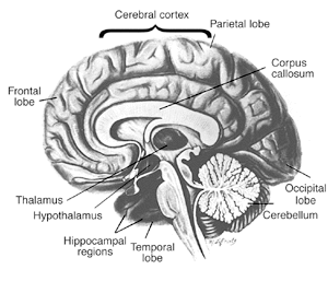
The cerebellum, an area of the brain responsible for coordinating movement and perhaps even some forms of learning, appears to be particularly sensitive to the effects of thiamine deficiency and is the region most frequently damaged in association with chronic alcohol consumption. Administering thiamine helps to improve brain function, especially in patients in the early stages of WKS. When damage to the brain is more severe, the course of care shifts from treatment to providing support to the patient and his or her family (18). Custodial care may be necessary for the 25 percent of patients who have permanent brain damage and significant loss of cognitive skills (19).

Scientists believe that a genetic variation could be one explanation for why only some alcoholics with thiamine deficiency go on to develop severe conditions such as WKS, but additional studies are necessary to clarify how genetic variants might cause some people to be more vulnerable to WKS than others.

LIVER DISEASE

Most people realize that heavy, long–term drinking can damage the liver, the organ chiefly responsible for breaking down alcohol into harmless byproducts and clearing it from the body. But people may not be aware that prolonged liver dysfunction, such as liver cirrhosis resulting from excessive alcohol consumption, can harm the brain, leading to a serious and potentially fatal brain disorder known as hepatic encephalopathy (20).

Hepatic encephalopathy can cause changes in sleep patterns, mood, and personality; psychiatric conditions such as anxiety and depression; severe cognitive effects such as shortened attention span; and problems with coordination such as a flapping or shaking of the hands (called asterixis). In the most serious cases, patients may slip into a coma (i.e., hepatic coma), which can be fatal.

New imaging techniques have enabled researchers to study specific brain regions in patients with alcoholic liver disease, giving them a better understanding of how hepatic encephalopathy develops. These studies have confirmed that at least two toxic substances, ammonia and manganese, have a role in the development of hepatic encephalopathy. Alcohol–damaged liver cells allow excess amounts of these harmful byproducts to enter the brain, thus harming brain cells.

Treatment

Physicians typically use the following strategies to prevent or treat the development of hepatic encephalopathy.

  • Treatment that lowers blood ammonia concentrations, such as administering L–ornithine L–aspartate.
  • Techniques such as liver–assist devices, or “artificial livers,” that clear the patients’ blood of harmful toxins. In initial studies, patients using these devices showed lower amounts of ammonia circulating in their blood, and their encephalopathy became less severe (21).
  • Liver transplantation, an approach that is widely used in alcoholic cirrhotic patients with severe (i.e., end–stage) chronic liver failure. In general, implantation of a new liver results in significant improvements in cognitive function in these patients (22) and lowers their levels of ammonia and manganese (23).

ALCOHOL AND THE DEVELOPING BRAIN

Drinking during pregnancy can lead to a range of physical, learning, and behavioral effects in the developing brain, the most serious of which is a collection of symptoms known as fetal alcohol syndrome (FAS). Children with FAS may have distinct facial features (see illustration). FAS infants also are markedly smaller than average. Their brains may have less volume (i.e., microencephaly). And they may have fewer numbers of brain cells (i.e., neurons) or fewer neurons that are able to function correctly, leading to long–term problems in learning and behavior.

Fetal Alcohol Syndrome

Treatment

Scientists are investigating the use of complex motor training and medications to prevent or reverse the alcohol–related brain damage found in people prenatally exposed to alcohol (24). In a study using rats, Klintsova and colleagues (25) used an obstacle course to teach complex motor skills, and this skills training led to a re–organization in the adult rats’ brains (i.e., cerebellum), enabling them to overcome the effects of the prenatal alcohol exposure. These findings have important therapeutic implications, suggesting that complex rehabilitative motor training can improve motor performance of children, or even adults, with FAS.

Scientists also are looking at the possibility of developing medications that can help alleviate or prevent brain damage, such as that associated with FAS. Studies using animals have yielded encouraging results for treatments using antioxidant therapy and vitamin E. Other preventive therapies showing promise in animal studies include 1–octanol, which ironically is an alcohol itself. Treatment with l–octanol significantly reduced the severity of alcohol’s effects on developing mouse embryos (26). Two molecules associated with normal development (i.e., NAP and SAL) have been found to protect nerve cells against a variety of toxins in much the same way that octanol does (27). And a compound (MK–801) that blocks a key brain chemical associated with alcohol withdrawal (i.e., glutamate) also is being studied. MK–801 reversed a specific learning impairment that resulted from early postnatal alcohol exposure (28).

Though these compounds were effective in animals, the positive results cited here may or may not translate to humans. Not drinking during pregnancy is the best form of prevention; FAS remains the leading preventable birth defect in the United States today.

GROWING NEW BRAIN CELLS

For decades scientists believed that the number of nerve cells in the adult brain was fixed early in life. If brain damage occurred, then, the best way to treat it was by strengthening the existing neurons, as new ones could not be added. In the 1960s, however, researchers found that new neurons are indeed generated in adulthood—a process called neurogenesis (29). These new cells originate from stem cells, which are cells that can divide indefinitely, renew themselves, and give rise to a variety of cell types. The discovery of brain stem cells and adult neurogenesis provides a new way of approaching the problem of alcohol–related changes in the brain and may lead to a clearer understanding of how best to treat and cure alcoholism (30).

For example, studies with animals show that high doses of alcohol lead to a disruption in the growth of new brain cells; scientists believe it may be this lack of new growth that results in the long–term deficits found in key areas of the brain (such as hippocampal structure and function) (31,32). Understanding how alcohol interacts with brain stem cells and what happens to these cells in alcoholics is the first step in establishing whether the use of stem cell therapies is an option for treatment (33).

SUMMARY

Alcoholics are not all alike. They experience different degrees of impairment, and the disease has different origins for different people. Consequently, researchers have not found conclusive evidence that any one variable is solely responsible for the brain deficits found in alcoholics. Characterizing what makes some alcoholics vulnerable to brain damage whereas others are not remains the subject of active research (34).

The good news is that most alcoholics with cognitive impairment show at least some improvement in brain structure and functioning within a year of abstinence, though some people take much longer (35–37). Clinicians must consider a variety of treatment methods to help people stop drinking and to recover from alcohol–related brain impairments, and tailor these treatments to the individual patient.

Advanced technology will have an important role in developing these therapies. Clinicians can use brain–imaging techniques to monitor the course and success of treatment, because imaging can reveal structural, functional, and biochemical changes in living patients over time. Promising new medications also are in the early stages of development, as researchers strive to design therapies that can help prevent alcohol’s harmful effects and promote the growth of new brain cells to take the place of those that have been damaged by alcohol.


Source: https://pubs.niaaa.nih.gov/publications/aa63/aa63.htm

References

(1) Parsons, O.A. Alcohol abuse and alcoholism. In: Nixon, S.J., ed. Neuropsychology for Clinical Practice. Washington, DC: American Psychological Press, 1996. pp. 175–201. (2) White, A.M. What happened? Alcohol, memory blackouts, and the brain. Alcohol Research & Health 27(2):186–196, 2003. (3) White, A.M.; Jamieson–Drake, D.W.; and Swartzwelder, H.S. Prevalence and correlates of alcohol–induced blackouts among college students: Results of an e–mail survey. Journal of American College Health 51:117–131, 2002. (4) Mumenthaler, M.S.; Taylor, J.L.; O’Hara, R.; et al. Gender differences in moderate drinking effects. Alcohol Research & Health 23:55–64, 1999. (5) Loft, S.; Olesen, K.L.; and Dossing, M. Increased susceptibility to liver disease in relation to alcohol consumption in women. Scandinavian Journal of Gastroenterology 22: 1251–1256, 1987. (6) Fernandez– Sola, J.; Estruch, R.; Nicolas, J.M.; et al. Comparison of alcoholic cardiomyopathy in women versus men. American Journal of Cardiology 80:481–485, 1997. (7) Ammendola, A.; Gemini, D.; Iannacone, S.; et al. Gender and peripheral neuropathy in chronic alcoholism: A clinical–electroneurographic study. Alcohol and Alcoholism35:368–371, 2000. (8) Jacobson, R. The contributions of sex and drinking history to the CT brain scan changes in alcoholics. Psychological Medicine 16:547–559, 1986. (9) Mann, K.; Batra, A.; Gunther, A.; and Schroth, G. Do women develop alcoholic brain damage more readily than men? Alcoholism: Clinical and Experimental Research16(6):1052–1056, 1992. (10) Nixon, S.; Tivis, R.; and Parsons, O. Behavioral dysfunction and cognitive efficiency in male and female alcoholics. Alcoholism: Clinical and Experimental Research 19(3):577–581, 1995. (11) Hommer, D.W. Male and female sensitivity to alcohol–induced brain damage. Alcohol Research & Health 27(2):181–185, 2003. (12) Hommer, D.W.; Momenan, R.; Kaiser, E.; and Rawlings, R.R. Evidence for a gender–related effect of alcoholism on brain volumes. American Journal of Psychiatry 158:198–204, 2001. (13) Pfefferbaum, A.; Rosenbloom, M.; Deshmukh, A.; and Sullivan, E. Sex differences in the effects of alcohol on brain structure. American Journal of Psychiatry 158:188–197, 2001. (14) National Academy of Sciences. Dietary reference intakes for thiamin, riboflavin, niacin, vitamin B6, folate, vitamin B12, pantothenic acid, biotin, and choline. 1999. (15) Morgan, M.Y. Alcohol and nutrition. British Medical Bulletins 38:21–29, 1982. (16) Martin, P.R.; Singleton, C.K.; and Hiller–Sturmhöfel, S.H. The role of thiamine in alcoholic brain disease. Alcohol Research & Health 27(2):134–142, 2003. (17) Victor, M.; Davis, R.D.; and Collins, G.H. The Wernicke–Korsakoff Syndrome and Related Neurologic Disorders Due to Alcoholism and Malnutrition. Philadelphia: F.A. Davis, 1989. (18) Martin, P. “Wernicke–Korsakoff syndrome: Alcohol–related dementia.” Family Caregiver Alliance Fact Sheet, 1998. (19) Cook, C. “The Wernicke–Korsakoff syndrome can be treated.” The Medical Council on Alcohol, vol. 19, 2000. (20) Butterworth, R.F. Hepatic encephalopathy—A serious complication of alcoholic liver disease. Alcohol Research & Health 27(2):143–145, 2003. (21) Mitzner, S.R., and Williams, R. Albumin dialysis MARS 2003. Liver International 23(Suppl. 3):1–72, 2003. (22) Arria, A.M.; Tarter, R.E.; Starzl, T.E.; and Van Thiel, D.H. Improvement in cognitive functioning of alcoholics following orthotopic liver transplantation. Alcoholism: Clinical and Experimental Research 15(6):956–962, 1991. (23) Pujol, A.; Pujol, J.; Graus, F.; et al. Hyperintense globus pallidus on T1–weighted MRI in cirrhotic patients is associated with severity of liver failure. Neurology 43:65–69, 1993. (24) Chen, W–J.A.; Maier, S.E.; Parnell, S.E.; and West, J.E. Alcohol and the developing brain: Neuroanatomical studies. Alcohol Research & Health 27(2):174–180, 2003. (25) Klintsova, A.Y.; Scamra, C.; Hoffman, M.; et al. Therapeutic effects of complex motor training on motor performance deficits induced by neonatal binge–like alcohol exposure in rats: II. A quantitative stereological study of synaptic plasticity in female rat cerebellum. Brain Research 937:83–93, 2002. (26) Chen, S.Y.; Wilkemeyer, M.F.; Sulik, K.K.; and Charness, M.E. Octanol antagonism of ethanol teratogenesis. FASEB Journal 15:1649–1651, 2001. (27) Spong, C.Y.; Abebe, D.T.; Gozes, I.; et al. Prevention of fetal demise and growth restriction in a mouse model of fetal alcohol syndrome. Journal of Pharmacology and Experimental Therapeutics 297:774–779, 2001. (28) Thomas, J.D.; Fleming, S.L.; and Riley, E.P. Administration of low doses of MK–801 during ethanol withdrawal in the developing rat pup attenuates alcohol’s teratogenic effects. Alcoholism: Clinical and Experimental Research26(8):1307–1313, 2002. (29) Altman, J., and Das, G.D. Autoradiographic and histological evidence of postnatal hippocampal neurogenesis in rats. Journal of Comparative Neurology 124(3):319–335, 1965. (30) Crews, F.T., and Nixon, K. Alcohol, neural stem cells, and adult neurogenesis. Alcohol Research & Health 27(2): 197–204, 2003. (31) Nixon, K., and Crews, F.T. Binge ethanol exposure decreases neurogenesis in adult rat hippocampus. Journal of Neurochemistry 83(5):1087–1093, 2002. (32) Herrera, D.G.; Yague, A.G.; Johnsen–Soriano, S.; et al. Selective impairment of hippocampal neurogenesis by chronic alcoholism: Protective effects of an antioxidant. Proceedings of the National Academy of Science of the U.S.A. 100(13):7919–7924, 2003. (33) Crews, F.T.; Miller, M.W.; Ma, W.; et al. Neural stem cells and alcohol. Alcoholism: Clinical and Experimental Research 27(2):324–335, 2003. (34) Oscar–Berman, M., and Marinkovic, K. Alcoholism and the brain: An overview. Alcohol Research & Health 27(2):125–133, 2003. (35) Bates, M.E.; Bowden, S.C.; and Barry, D. Neurocognitive impairment associated with alcohol use disorders: Implications for treatment. Experimental and Clinical Psychopharmacology 10(3):193–212, 2002. (36) Gansler, D.A.; Harris, G.J.; Oscar–Berman, M.; et al. Hypoperfusion of inferior frontal brain regions in abstinent alcoholics: A pilot SPECT study. Journal of Studies on Alcohol 61:32–37, 2000. (37) Sullivan, E.V.; Rosenbloom, M.J.; Lim, K.O.; and Pfefferbaum, A. Longitudinal changes in cognition, gait, and balance in abstinent and relapsed alcoholic men: Relationships to changes in brain structure. Neuropsychology 14:178–188, 2000. (38) Rosenbloom, M.; Sullivan, E.V.; and Pfefferbaum, A. Using magnetic resonance imaging and diffusion tensor imaging to assess brain damage in alcoholics. Alcohol Research & Health 27(2):146–152, 2003. (39) Kensinger, E.A.; Clarke, R.J.; and Corkin, S. What neural correlates underlie successful encoding and retrieval? A functional magnetic resonance imaging study using a divided attention paradigm. Journal of Neuroscience 23(6):2407–2415, 2003. (40) Wong, D.F.; Maini, A.; Rousset, O.G.; and Brasíc , J.R. Positron emission tomography—A tool for identifying the effects of alcohol dependence on the brain. Alcohol Research & Health 27(2):161–173, 2003. (41) Porjesz, B., and Begleiter, H. Alcoholism and human electrophysiology. Alcohol Research & Health 27(2):153–160, 2003. (42) Porjesz, B., and Begleiter, H. Human brain electrophysiology and alcoholism. In: Tarter, R., and Van Thiel, D., eds. Alcohol and the Brain. New York: Plenum, 1985. pp. 139–182. (43) Begleiter, H.; Porjesz, B.; Bihari, B.; and Kissin, B. Event–related potentials in boys at risk for alcoholism. Science 225:1493–1496, 1984. (44) Polich, J.; Pollock, V.E.; and Bloom, F.E. Meta–analysis of P300 amplitude from males at risk for alcoholism. Psychological Bulletin 115:55–73, 1994.


Anti-Seizure Effects of the Ketogenic Diet

The Gut Microbiota Mediates the Anti-Seizure Effects of the Ketogenic Diet

Christine A. Olson, Helen E. Vuong, Jessica M. Yano, Qingxing Y. Liang, David J. Nusbaum, Elaine Y. Hsiao2, Correspondence information about the author Elaine Y. HsiaoEmail the author Elaine Y. Hsiao
2Lead Contact

Highlights

  • Changes in the gut microbiota are required for the anti-seizure effects of the KD
  • Specific KD-associated bacteria mediate and confer the anti-seizure effects of the KD
  • KD microbiota regulate amino acid γ-glutamylation and hippocampal GABA/glutamate

Summary

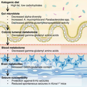
The ketogenic diet (KD) is used to treat refractory epilepsy, but the mechanisms underlying its neuroprotective effects remain unclear. Here, we show that the gut microbiota is altered by the KD and required for protection against acute electrically induced seizures and spontaneous tonic-clonic seizures in two mouse models. Mice treated with antibiotics or reared germ free are resistant to KD-mediated seizure protection. Enrichment of, and gnotobiotic co-colonization with, KD-associated Akkermansia and Parabacteroides restores seizure protection. Moreover, transplantation of the KD gut microbiota and treatment with Akkermansia and Parabacteroides each confer seizure protection to mice fed a control diet. Alterations in colonic lumenal, serum, and hippocampal metabolomic profiles correlate with seizure protection, including reductions in systemic gamma-glutamylated amino acids and elevated hippocampal GABA/glutamate levels. Bacterial cross-feeding decreases gamma-glutamyltranspeptidase activity, and inhibiting gamma-glutamylation promotes seizure protection in vivo. Overall, this study reveals that the gut microbiota modulates host metabolism and seizure susceptibility in mice.

Night owls die young in a world scheduled for early birds

Living out of sync with the rest of the world may be to blame

by Maggie Fox and Erika Edwards / 

The stress of night owls trying to live against their nature could be a factor in their increased risk of dying over a given period of time.Luciano Lozano / Getty Images/Ikon Images

Are you a night owl? It might be killing you, researchers reported Thursday.

They found people with naturally late body clocks were about 10 percent more likely to die over a given period than early birds who rise with the sun.

It’s probably because living in a world geared for early starts is throwing off the circadian rhythms of the night owls, the researchers said.

Their findings fit in with other reports that show people who stay up later at night have higher risks of diabetes, high blood pressure and some types of cancer.

The researchers looked at surveys of more than 400,000 people taking part in a large British study of genes and health. As part of a detailed questionnaire, they were asked whether they tended to be night owls or morning larks.

There wasn’t much difference among people who fell in the middle. But there was a notable difference between the two extremes, said Kristen Knutson, associate professor of neurology at Northwestern University Feinberg School of Medicine.

“We found that the night owls had a 10 percent increased risk of dying over about a six and a half year period. And that was even after we took into account things like existing health problems,” Knutson told NBC News.

It’s not a lack of sleep — both groups got about the same amount, Knutson and her colleague Malcolm von Schantz, a professor of chronobiology at the University of Surrey, reported in the journal Chronobiology International.

“I think the problem arises because a night owl is trying to live in a morning lark world,” Knutson said. “If the body is expecting you to do something at a certain time like sleep or eat and you’re doing it at the quote ‘wrong’ time, then your body’s physiology may not be working as well.”

Researchers know the body clock is important. Three scientists who study the body’s internal clock won the Nobel Prize in Medicine last year. Their work, done over decades, helps explain how life adapts to the 24-hour cycle of day, and also how diseases such as cancer arise in the cells.

Studies indicate that switches to and from daylight savings time can raise the immediate risk of death over the following days, and others show the health risks of shift work.

The International Agency for Research on Cancer, an arm of the World Health Organization, says shift work probably causes cancer. It’s linked with breast cancer and other types of cancers, as well as diabetes and sleep disruption.

Those studies support the idea that working against the body’s natural inclination can be hazardous to your health.

Knutson and Von Schantz looked at how people answered the early bird question. “Approximately, 27 percent identified as definite morning types, 35 percent as moderate morning types, 28 percent as moderate evening types and 9 percent as definite evening types,” they wrote.

It was the 9 percent of people who said they were “definite” evening types who had the 10 percent increase risk of dying from something over the next six or so years.

“Neither of the two intermediate groups was associated with increased risk of all cause mortality,” they added.

There could be many reasons. People who stay up later eat fattier foods, drink more alcohol and are more likely to use recreational drugs. They have more exposure to artificial light, as well.

“Greater eveningness has also been associated with depression and mood disorders, particularly in those 50 years or older,” the researchers added.

But the stress of trying to live against their natures could be at fault, they proposed.

“The health of evening types could be compromised by misalignment between their endogenous biological clocks and the timing of social activities (e.g. work or meals), termed circadian misalignment,” they wrote.

WHAT CAN PEOPLE DO?

“You can’t just suddenly go to bed three hours earlier tonight because it won’t work,” Knutson said.

Confirmed night owls must make sure to eat a healthy diet and exercise regularly, she said.

But society should also recognize that some people have a genetic tendency to sleep later.

“If we can recognize these chronotypes are, in part, genetically determined and not just a character flaw, jobs and work hours could have more flexibility for owls,” Knutson said.

“This is a public health issue that can no longer be ignored,” von Schantz added.

“We should discuss allowing evening types to start and finish work later, where practical. And we need more research about how we can help evening types cope with the higher effort of keeping their body clock in synchrony with sun time.”